 English




Histology and Anatomy Laboratory
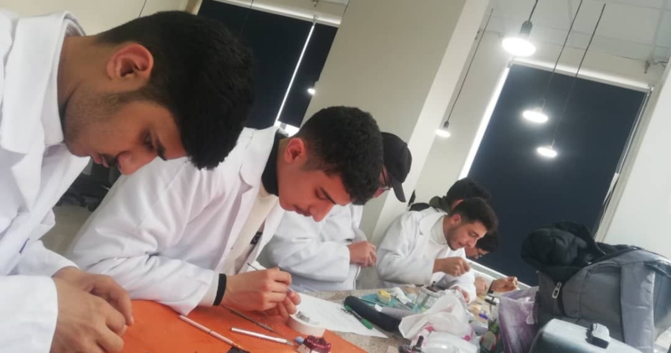
The Histology and Microscopic Anatomy Laboratory at the Faculty of Dentistry, Qasyoun Private University, provides a rigorous scientific foundation for understanding the microscopic and fine-grained structure of human tissues in general, and oral and dental tissues in particular, linking the external appearance of organs to their fine cellular composition.
General Objectives of the Laboratory:
Understanding Cellular and Histological Structure: Studying the four basic tissues of the body (epithelial, connective, muscular, and nervous).
Deep-seated Oral and Dental Histology: Comprehending the microscopic and fine-grained structure of enamel, dentin, cementum, dental pulp, and supporting tissues (periodontal ligament, alveolar bone, and gingiva).
Distinguishing Between Normal and Pathological Conditions: Enabling students to identify healthy tissue structures, which will serve as a fundamental reference in diagnosing pathological changes (oral pathology) later on.
Available Equipment and Technologies:
Modern Light Microscopes: Binocular microscopes with varying magnification powers allow students to examine tissue slides with high precision. Digital Microscope Systems: Devices for displaying tissue slides directly on interactive screens for group explanations and discussions.
Prepared Histological Slides: Sets of colored, pre-prepared histological slides covering all body organs and oral tissues in their various developmental stages.
Odontogenesis Models: Models illustrating the stages of tooth bud formation and differentiating dental tissues.
Student Training Topics and Skills:
General Histology:
Study of different types of epithelium, connective tissues, and skeletal tissues.
Examination of histological sections of blood vessels, nerves, and various types of muscles.
Oral Histology:
Hard Dental Tissues: Study of the microscopic structure of enamel rods, dentin tubules, and cementum.
Pulp and Supporting Tissues: Examination of odontoblasts, alveolar ligament fibers, and the cellular structure of the gingiva. Salivary Glands and Oral Mucosa: Study of histological sections of the major and minor salivary glands and the oral mucosa.
Tooth Development and Formation (Odontogenesis): Monitoring the stages of tooth bud formation (Bud, Cap, and Bell Stages) and the stages of calcification and root development.
The Importance of the Laboratory in Clinical Practice: This laboratory forms the cornerstone for understanding oral pathology, endodontics, and oral and maxillofacial surgery. Drug response, surgical healing, and the mechanisms of caries and bone resorption all depend on a deep understanding of the cellular and histological structures covered by this laboratory.