 English

X-ray laboratory
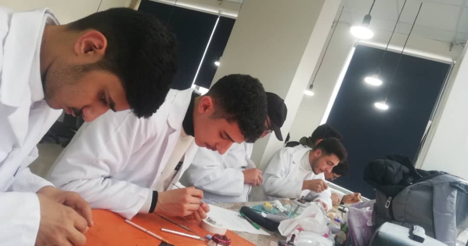
The Radiology Laboratory at the Faculty of Dentistry, Qasyoun Private University, provides students with practical and applied training in various dental radiography techniques. It is one of the most important diagnostic laboratories, enabling students to visualize structures invisible to the naked eye within the jaws and skull.
General Objectives of the Laboratory:
Understanding Radiologic Physics and Imaging Mechanisms: Studying the mechanism of X-ray generation and their interaction with dental and bone tissues.
Mastering Intraoral and Extraoral Imaging Techniques: Training students on patient positioning, X-ray tube angles, and film supports to obtain high-resolution diagnostic images.
Reading and Analyzing Radiographic Images: Developing the skill of interpreting radiographic shadows, distinguishing normal from abnormal tissues, and detecting dental and bone lesions.
Applying Radiological Safety: Establishing the principles of ALARA (Accessible Radiological Protection) to protect patients, dentists, and technical staff from the dangers of cumulative radiation exposure. Available Equipment and Technologies
Intraoral X-ray Units: Wall-mounted and mobile units designed for periapical, pterygoid, and occipital imaging.
Digital Radiovisual (RVG) Systems: Digital sensors and probes connected to a computer to produce images instantly with very low radiation doses compared to conventional techniques.
Panorama/Orthopantomogram Units: Extraoral units providing a complete and comprehensive scan of the jaws, teeth, temporomandibular joint (TMJ), and maxillary sinus.
Film Holders & Positioners: Instruments for adjusting the paralleling technique and the bisecting angle technique.
Radiation Protection Equipment: Lead aprons, thyroid shields, and lead-lined chambers/doors to shield the area from radiation. Student Training Topics and Skills
Intraoral Imaging Techniques:
Apical technique: To visualize the entire tooth from the crown to the root apex and surrounding supporting tissues.
Periodontical technique: To detect interdental caries and assess the level of residual bone.
Extraoral Imaging Techniques and Interpretation:
Studying panoramic images and analyzing the impaction path of teeth (especially wisdom teeth), fractures, and bone lesions.
Radiological Diagnosis and Interpretation:
Distinguishing carious lesions, bone resorption, cysts, and tumors.
Interpreting endodontic treatment images (root canal fillings) and confirming that the filling reaches the apex.
Radiation Safety and Protection Measures:
Applying the ALARA principle (minimizing radiation dose to the lowest reasonably achievable level).
Optimizing the use of lead shielding and adjusting the exposure time appropriately according to the patient's age and anatomy. The importance of the laboratory in the clinical process
The radiology laboratory is a pivotal step in preparing the dentist, as no root canal, surgical, orthodontic, or dental implant treatment can be performed without complete reliance on accurate radiographic diagnosis to prepare the appropriate treatment plan.